

CBCT Scan in Dentistry: What Can It Reveal That a Regular X-Ray Cannot?

Dr. D.Sendhil Nathan
MDS, DNB (Prosthodontics And Implantology)
Sometimes a dental X-ray gives the dentist all the information needed. But there are situations where looking at a tooth from just one angle is not enough. The dentist may need to understand the depth of the jawbone, the exact position of a tooth, or how a tooth relates to nearby structures.
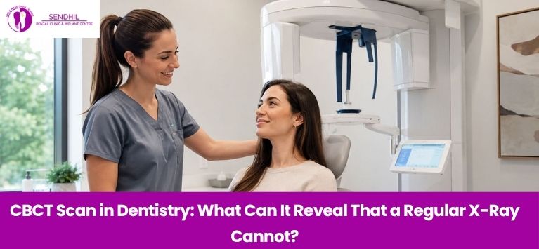
This is where a CBCT scan in dentistry can be useful. CBCT, or Cone Beam Computed Tomography, produces a three-dimensional image of the teeth, jaws and surrounding areas. Instead of relying only on a flat image, the dentist can examine the scanned area from different directions.
Table of Contents
What Makes a CBCT Scan Different?
A conventional dental X-ray creates a two-dimensional picture. It is extremely useful for everyday dental examinations, but structures that sit at different depths can appear superimposed on one another.
CBCT works differently. The scanner captures multiple X-ray images while rotating around the patient’s head. A computer then processes these images to create a 3D view of the area being examined.
Think of it this way: a regular X-ray gives your dentist a picture, while CBCT can provide a much clearer sense of where things are in three dimensions.
That extra information is not necessary for every patient. CBCT is generally considered when the additional detail could influence diagnosis or treatment planning.
What Can CBCT Show More Clearly?
The biggest advantage of CBCT is the ability to assess the relationship between different structures rather than viewing them as overlapping shapes.
Depending on the reason for the scan, it may help the dentist examine:
- The height and width of the jawbone
- The position and orientation of impacted teeth
- The shape and location of tooth roots
- The relationship between teeth and surrounding structures
- The available bone at an implant site
- Certain changes affecting the jaw or facial structures
- Areas that need closer assessment before surgery
This three-dimensional information can be particularly valuable when a treatment requires careful planning.
Why Is CBCT Important Before Dental Implants?
Dental implant placement involves more than checking whether a space is available for a missing tooth. The dentist also needs to understand the bone surrounding the proposed implant site and its relationship with important anatomical structures.
CBCT can provide cross-sectional information about the implant area, helping the dental team assess the available bone and plan the position of the implant.
For more complex implant treatments, this information can be combined with digital planning tools to create a more detailed treatment plan.
Sendhil Dental’s implant treatment information describes digital planning with CBCT of the jaws as part of its implant assessment process.
Can CBCT Help With Wisdom Teeth?
It can be useful when a wisdom tooth has an unusual position or when its relationship with surrounding structures needs to be understood more clearly.
A regular X-ray may show that a wisdom tooth is impacted, but a 3D scan can provide additional information about its orientation and position within the jaw.
This can be helpful when the dentist is planning treatment for a complicated impaction. It allows the area to be viewed from multiple directions instead of relying on a single two-dimensional projection.
CBCT and Regular X-Ray: Are They the Same?
CBCT Scan | Regular Dental X-Ray |
| Produces a 3D image | Produces a 2D image |
| Useful when additional 3D detail is required | Useful for many routine examinations |
| Allows examination from different planes | Structures may overlap |
| Particularly useful for complex treatment planning | Often sufficient for straightforward cases |
Generally involves more radiation than conventional dental X-rays | Lower radiation exposure than CBCT |
Is a Dental CBCT Scan Safe?
CBCT uses X-rays and therefore involves exposure to ionising radiation. The radiation dose varies according to the equipment, settings and size of the area being scanned.
For this reason, CBCT should be used when the expected diagnostic benefit justifies the exposure. The FDA and ADA recommend that dental imaging, including CBCT, should be performed when it is clinically necessary rather than simply as a routine procedure.
If your dentist recommends a CBCT scan, you can ask what information they need from it and how it will affect your treatment plan.
When Might Your Dentist Recommend CBCT?
CBCT may be considered when detailed 3D information is useful for:
- Dental implant planning
- Complex or impacted wisdom teeth
- Selected root canal cases
- Oral surgery planning
- Assessment of complicated tooth or jaw anatomy
- Certain dental injuries or bone-related conditions
Final Thoughts
Regular dental X-rays remain an important part of everyday dental care, but they may not provide enough detail for every situation. When a dentist needs to understand the depth, position, and relationship between different structures, CBCT can offer a more detailed 3D view.
A CBCT scan in dentistry can be particularly useful when detailed planning is needed for dental implants and certain complex dental procedures. At Sendhil Dental, CBCT forms part of the digital planning process used for implant treatment, helping the dental team assess each case in greater detail.